Table of Contents
The Hidden Engines of Research
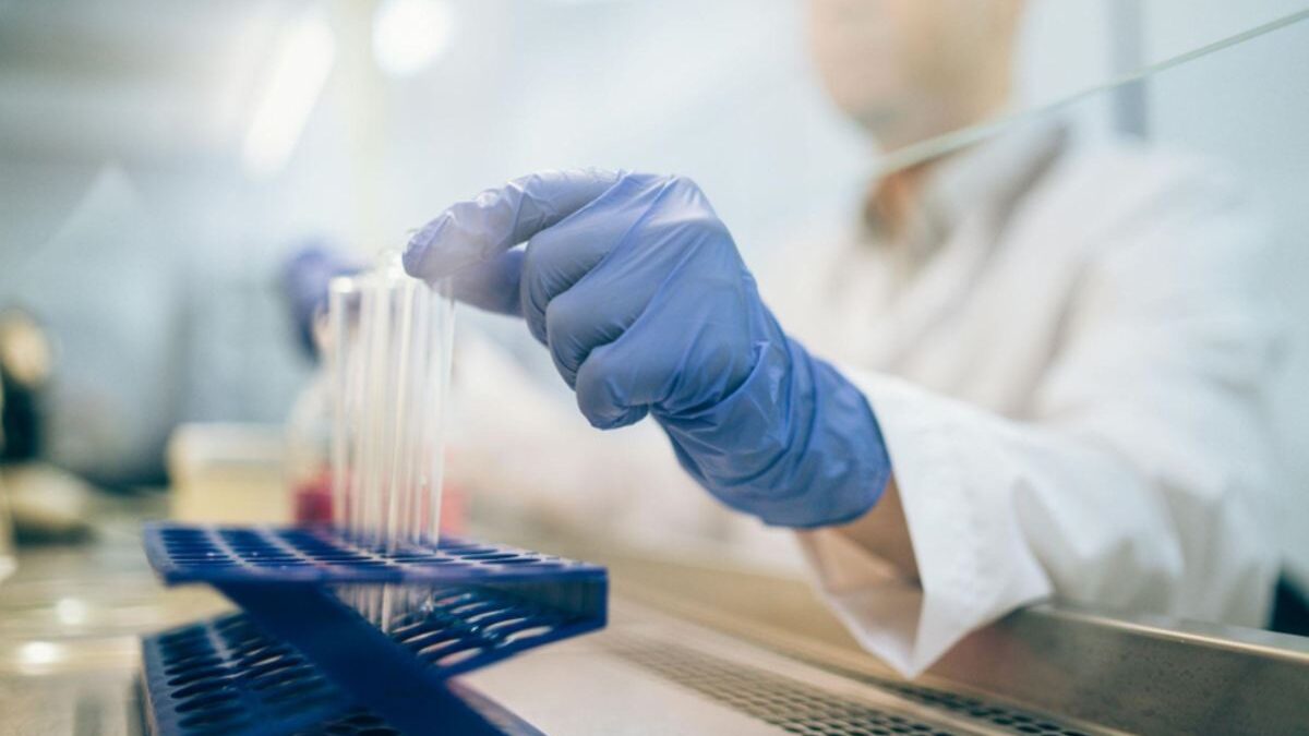
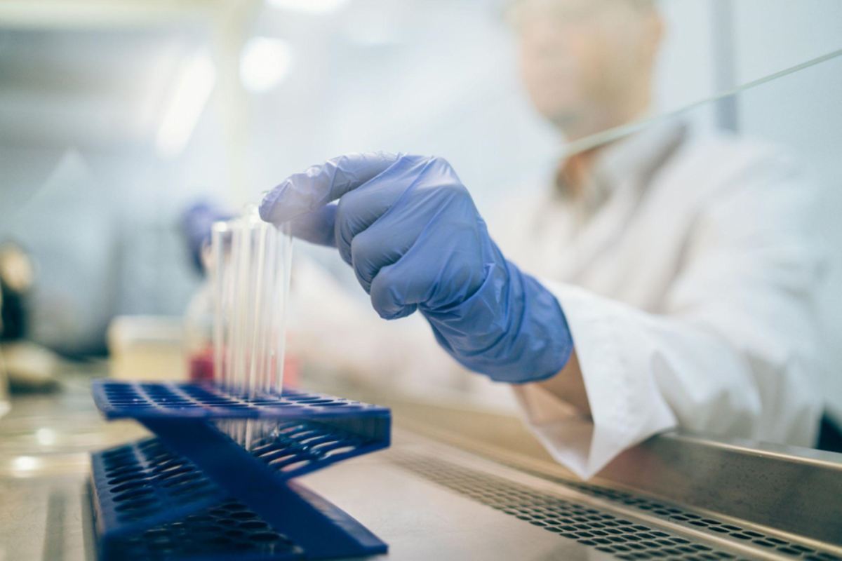
Biotech labs feel like a mix of science and creativity. Researchers spend long hours studying cells and processes that we rarely see. The work shapes medicine, therapies, and even how we understand the body. Many of these advances come from tools that are not often discussed outside lab walls.
One of those tools is the live cell imager. It is not just another piece of equipment. It is a window into the life of cells as they grow, react, and change. This machine helps researchers watch biology happen in real time.
What a Live Cell Imager Actually Does
At its core, the live cell imager records cell activity while keeping cells alive. Traditional microscopes can show cells, but they often require fixed samples. That means the cells are not active anymore. A live cell system solves that problem.
The machine maintains the right conditions for the cells. It controls temperature, gas levels, and moisture. It also uses advanced optics and cameras to capture images at set intervals. This allows scientists to watch changes over minutes, hours, or even days.
The result is a moving record of cell behavior. That kind of view offers insight that still images can never provide.
Why Real-Time Imaging Matters
Cells are dynamic. They move, divide, and respond to signals from their environment. A snapshot can miss critical steps. With live imaging, every stage becomes visible.
This helps researchers track growth patterns. It allows them to see how cells respond to treatments. It shows interactions that shape larger biological systems. For drug development, this can be a game-changer. It allows scientists to spot effects quickly and adjust their studies with more precision.
Real-time imaging also boosts confidence in results. Instead of guessing what happened between two static points, researchers can see the full timeline.
The Technology Behind the Lens
A live cell imager is more than a camera. It is a system built from multiple technologies working together. High-resolution optics provide clear detail. Automated stages move plates or slides with accuracy. Advanced software runs the imaging cycles and manages the data.
Many systems use fluorescence to highlight structures or proteins. This makes certain features stand out against the cell background. The software can then analyze those signals, track them, and present usable data.
Every piece is designed for one goal: capturing the most accurate view of living cells without disturbing them.
Integration With Lab Workflows
Modern labs depend on efficiency. Adding new equipment only works if it fits smoothly into daily routines. Live cell imagers are designed with that in mind.
They connect with standard lab plates and formats. They often link with analysis software used across other machines. Many can run automated programs, which means staff can set them up and let them run while focusing on other work.
This integration is key for high-volume labs. It reduces wasted time and ensures that the imager supports, rather than slows down, ongoing research.
From Research to Real-World Impact
The images captured in a lab are not just for curiosity. They form the base of discoveries that may lead to new treatments and health solutions. Watching cells respond to new drugs can guide which compounds move forward. Seeing how cells interact can shape understanding of disease pathways.
This direct link between imaging and outcome is why live systems hold so much value. They make the invisible visible, and they do it in a way that drives practical progress.
In biotech, that connection between basic research and applied science is the heart of innovation.
Challenges and Care Needs
Like any advanced tool, live cell imagers require proper care. They must be calibrated and maintained. Staff need training to set up experiments and manage conditions. If the environment is not right, the cells can fail, and the results will lose value.
Data storage is another factor. The imager produces a large amount of visual information. Labs must plan for how to handle and archive those files. Many systems include built-in storage and analysis, but long-term data handling often needs extra planning.
These challenges do not reduce the value of the machine. They just highlight that it is a serious investment that requires planning and support.
Looking Ahead in Imaging Technology
Technology rarely stands still. Live cell imaging is already seeing upgrades in automation, resolution, and data processing. Some systems can now analyze results in real time while the experiment is still running. Others connect directly to cloud platforms for instant sharing.
The future will likely bring even smarter systems. Imagine imagers that adjust conditions based on cell behavior. Or tools that predict outcomes as they record data. These kinds of advances could make labs even more efficient and accurate.
For now, live cell imagers remain one of the most exciting tools in biotech labs. They capture life in action and turn it into knowledge. That knowledge drives discoveries that shape the future of medicine.